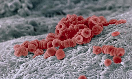
Red blood cells on an artery wall Photograph: Corbis Flirt / Alamy/Alamy
If you press firmly on one of your fingernails and then release the pressure, the nail-bed will blanch momentarily before pinking up again; it's a simple test used to assess perfusion – the extent to which tissues are adequately oxygenated. And it is the red blood cell that is responsible for this critical job.
When levels of oxygen in a person's tissue decrease – for example from exercise, trauma or disease – the kidney releases a hormone called erythropoietin. This stimulates the production of new red blood cells. Just as they are about to enter the circulation, the cells lose their nucleus making them incredibly light and bendy, and giving them their characteristic biconcave shape. By folding up like tiny umbrellas, they are now able to fit through the narrowest of capillaries, and so deliver essential oxygen.
Red blood cells contains large amounts of haemoglobin – the protein that gives blood both its colour and its oxygen-binding capacity. Each one lives for about a 120 days and travels an amazing 300 miles around the body